Human on Chip is Possible and its Coming

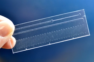
The sleek contraption that won this year’s UK Design Award doesn’t resemble a living organ. But this translucent, glowing microchip, with slender tubes branching off of it, breathes just like a human lung. The device, which was created by researchers at Harvard’s Wyss Institute for Biologically Inspired Engineering, is made from a rubber-like polymer and is a mixture of biological human cells and fluid that mimics blood. The lung device is just one of several “human organ simulators” designed by the researchers, Daniel Huh, a former Wyss Institute Fellow and assistant professor in bioengineering at the University of Pennsylvania, told me over the phone. “They are tiny microchips about the size of a computer memory stick that we use to grow living cells, and mimic the most important structure and function of human organs.” Huh and his colleagues have already made devices that mimic lungs, kidneys, and skin. “We’re doing many different organs and by having a common blood vessel channel, we can actually link them together to eventually create a human body on a chip,”Donald E. Ingber, bioengineer and Wyss’ Founding Director, told me. “We’ve already connected four different organ on chips together and kept them alive on an automated instrument for two weeks.” The researchers aim to use the devices to create a more holistic representation of the human body, making drug testing models cheaper, more ethical, and more effective. “It would allow us to mimic the human biology and physiology at the whole body level, not just at the individual level,” said Huh. “Our design was motivated by one of the most critical medical challenges that we’re facing: That the animal models that we use for developing and testing new drugs are for the most part failing to predict how humans respond to drugs,” he said. “The biology of animals like mice is completely different than that of humans.”

The lung on a chip was made using a microfabrication process adapted from the computer chip industry. It sandwiches two layers of living tissue composed of cells from the lung’s air sacs and the surrounding blood vessels across a porous rubber membrane containing hollow microchannels. A culture medium mimicking blood flows in the capillary channel, and a regular mechanical stretching function mimics breathing motions. Air and fluid are delivered via tubing connected to the microchip by using a syringe and vacuum pumps.

What’s exciting about the device is its ability to replicate complex biological processes. For example, when bacteria is placed on the surface of the chip to mimic a lung infection, and white blood cells taken from the human body are placed into the blood capillary channels of these devices, the chip reacts fiercely.

“The white blood cells circulating in the capillary channel stick to capillary cells, get across the tissue layer, show up on the lung surface and start engulfing the bacteria,” said Huh. “Just as they would do in a lung in the human body.”

Currently, drug developers either test drugs on animals or use a cell culture model where they take living cells from a human body, grow them in a petri dish, then test drugs on them. While animal testing evokes ethical issues, the petri dish approach hardly replicates all the complexities of the human body with its beating heart, contracting muscles, and constant blood flow.

“If you think about it, this is such an unnatural, uncomfortable environment for these cells. They are used to very complex, dynamic three-dimensional environments in the human body,” said Huh.

He explained that traditional drug testing methods are largely failing to predict the effects of drugs at a preclinical stage. This has a knock-on effect at the human trial stage as both time and money are ill-spent on drugs that end up having no impact.

“The idea here is by developing next-generation drug testing technologies we can use the preclinical stage to weed out the drugs that are destined to fail as early in the process as possible,” said Huh. This, he explained, would dramatically reduce both the cost and time required to develop effective drugs in the future.

An out of Body Experience Organ-On-Chips

Pioneering work in the United States to create organs-on-chips could revolutionize the future of drug development. Four years ago scientists observed how a white blood cell reacts when it senses an infection. They watched the leukocyte as it wriggled through capillary cells the cells that line blood vessels and then through the cells that line the lung, to then engulf an invading bacterium. But this was not happening inside a patient, it was happening on a microchip. “It mimics the human response, it’s amazing to watch,” says Geraldine Hamilton, senior staff scientist at the Wyss Institute for Biologically Inspired Engineering.

Researchers at the Wyss Institute, which is based at Harvard University in Boston, Massachusetts, are pioneering the development of a whole pipeline of human organs-on-chips, including the lung, gut, heart, liver, skin, bone marrow, pancreas, kidney, eye and even a system that mimics the blood-brain barrier. The idea is to recreate the smallest functional unit of any particular organ in a micro-environment that closely imitates the human body, explains Hamilton.

To the naked eye, these ‘organs’ look nothing like the human body. A clear, flexible polymer is used to form the rectangular chips, which are about the size of a memory stick. Tiny channels pass through the chips like miniature ribbons, lined with living human cells, which vary depending on the organ being modelled. Other elements of the chip can also be customised.

So, for example, to create a lung-on-chip, the channels are split horizontally down the middle with a porous membrane. On top of the membrane, human lung cells grow and below it grow capillary cells. Air is passed over the top of the lung tissue and blood cells flow underneath the layer of capillary cells — mimicking the interface between the alveoli, the pockets of lung into which air is inhaled, and the blood vessels that carry oxygen away, all within the same tiny channels.

The flexibility of the chips means the mechanical forces that cells experience inside the human body can also be recreated. In the case of the lung-on-chip, the motion of breathing is simulated by applying a vacuum to side channels on the left and right of the main channel, stretching and relaxing the lung tissue inside.

This is important. Hamilton explains that Donald Ingber, who leads the organ-on-chip work at Wyss, showed that mechanical forces are key drivers of how a cell functions.

“We are trying to create an environment where they can function like they would in the body,” says Hamilton. Organs-on-chips can be used to determine whether a particular protein is a suitable target for drug development, to identify drug toxicity or even to assess efficacy, says Hamilton.

The technology is still in its infancy, but there is a need for it; at the moment the techniques used to discover and develop drugs — animal models and human cell lines — are too often failing to predict what will happen in humans. Of the drugs that make it to phase I trials, only about 10% successfully reach the market.

Hamilton believes that organs-on-chips could revolutionize the pharmaceutical industry by making drug development faster and cheaper, and by producing more successful therapies. Different disease states can be modelled on the chips and, crucially, drugs can be added to the channels and their effects on the tissue examined.

The Origin of Microfuidics

The microfluidics based devices have intellectual roots in at least two diverse fields: microelectronics and bioanalytical chemistry. The computer chip paradigm inspired chemists and engineers to apply some of these same technologies to generate networks of channels through which fluids can be moved.

Not coincidentally, commercial microfluidics has its origins in California's Silicon Valley. The advent of microprocessors in the early 1970s and the subsequent development of highly integrated microelectronic devices later in the decade paved the way for microfluidics. The application of microfluidics to the bioanalytical realm derives from work done in the late 1980's by Andreas Manz, then an analytical chemist at Ciba Geigy (now Novartis), who originated the TAS , an early lab-on-a-chip. Manz envisioned the application of photolithography and chemical etching techniques for micromachining a complete microanalytical system on a chip to integrate sample preparation, chemical derivatization, electrophoretic separation, and detection using only nanoliters or picoliters of the test analyte.

In the course of investigating the TAS concept, Manz and his co-workers discovered that the phenomenon of electro-osmosis could be used to control the injection, flow and mixing of solutions.

Manz and coworkers joined forces with Jed Harrison's group (University of Alberta, Canada) to demonstrate the practicality of such a device. Their studies showed that channels 10m deep by 30m wide etched in glass could be used for capillary electrophoresis, offering high-resolution separations in as little as 15 seconds.

More importantly, they showed that creating electric potential differences across chip elements permitted the manipulation of flow and mixing of fluids. The combination of exceptionally fast separation times and miniscule sample sizes demonstrated that such micro-systems were not simply elegant curiosities, but had practical advantages as well.

The integration of nucleic acid sample preparation, amplification, and hybridization, became another technological goal for the microfluidics community during the 1990s.

The first major commercial thrust toward micro-scale nucleic acid target amplification originated from work done by Allen Northrup and co-workers (Lawrence Livermore Laboratory).2 They demonstrated that PCR reactions could run up to ten times faster in their micro-scale device than by conventional means.

Kricka and Wilding (University of Pennsylvania School of Medicine) independently devised a thermocycler capable of amplifying DNA contained in less than five microliters of solution.

In 1994, J. Michael Ramsey and co-workers (U.S. Department of Energy's Oak Ridge National Laboratory) began publishing a series of papers that extended the findings of Manz and Harrison and provided the fledgling microfluidics enterprise with some of its key intellectual property. The Ramsey group described a prototypical microfluidic device consisting of two perpendicular etched channels in the form of a cross with four reservoirs, one at each channel terminus.

Knowing About Microfluidic Chips

Although microfluidics has been a buzz word within industrial applications over the past 20 years, few developments in the point of care diagnostic sector have been transferred from the research setting to a commercial product that can be manufactured in high volume. The benefits of microfluidics are numerous: ease of use, reduced sample volumes and reaction times, higher data quality and reliable parameter control. The intricacy of the microfluidic chips means that little is required of the detection instrument and the integral automation means the user does not need to engage in multi-step reactions requiring a high level of skill. The challenge lies with validating the complex processes that deliver reagents in order, accurately and reproducibly.

Alongside the technical issues associated with microfluidic platforms the diagnostics industry also has difficulty in identifying a low cost manufacturing technique for the micro patterning needed. This article will explore these challenges and offer potential solutions.

Whilst the paramount requirement is result accuracy, any device designed for point of care also needs to be small, cheap, mobile, robust and fast to be successful. Microfluidic techniques allow manufacturers to produce devices which are lower cost, smaller and potentially hand-held. At the same time, all of the profit of a microfluidic diagnostic kit sits with the consumable device, so manufacturers need to focus on keeping unit costs low while maintaining efficacy.

‘Microfluidics as a basic processing technology clearly works at the proof of principle level but that is where most of the work ends, usually in a publication. Engineering the device into a manufacturable product requires some consideration of substrate material, device functionalisation, packaging and performance testing. Currently the biggest opportunity and largest potential barrier to moving micro and nano technology into the commercial environment lies in innovative manufacturing’, comments key opinion leader on lab-on-a-chip and microfluidic technology Professor Steve Haswell, University of Hull.

For polymer devices, two leading technologies can be applied to enable cost-effective mass production: Injection Moulding (IM) and Polymer Laminate Technology (PLT).

IM carries many advantages in addition to its rapid manufacture: it is low cost with secure supply chains and a large palette of polymers available.

Mass production of microfluidic chips using IM is perceived to be problematic because it lacks the precision to reproduce micro features reliably due to effects of polymer creep or shrinkage in the final products.

Looking to the horizon of next generation microfluidic diagnostics, research has been focused on microfluidic devices being produced on paper. Analogous to lateral flow diagnostics, these microfluidic devices use hydrophobic channel walls printed on paper to allow the liquid flow direction and speed to be controlled. These paper based devices are extremely low cost and readily disposable. Although currently limited to relatively simple device architectures, this new platform approach is certainly one to watch for the future.

How online connects in Everyone’s Life

In the world of s science there are a lot of things that can’t be understand easily for the scientists that we have in this word they are still proving every theory that they have in order everyone will believe in science. Their worlds is way more complicated in the normal life that we have keeping us imagine how their brain works 24/7. Sounds really hilarious but what can we do it will make us thinks like that.

One of the fields that take a lot of time and effort with those brain storming activities in their field are in the medical. For my opinion people that are involved in the medical field especially for those in the research are the tough one’s imagine the time and effort that they take in order to gather and proves every thing that they do for the betterment of the society. It is not just hard but the word that fits is complicated. I’m must be thinking are they sleeping at night with the same amount of hours of the normal person off course they do not because they maybe sleeping in thier lab all the time. Well, in the part of the research that they have one of the critical part is when the fluids are included without the proper instrument to be used everything might goes wrong. Exact measurement is very important all the time that is why most of them are using the appropriate instruments.

Every instrument is just design for their perfect usage and also the manufacturer always looks out for it. So where you can find these kinds of instruments like fluid control pumps? Today online is the best source of everything even in shopping do it online is one of the convenient way in looking for the specific item for a specific things. One of the newly firm online that meets in your needs when it comes to fluid control products is Corsolutions. CorSolutions creates and commercializes innovative, intuitive products that support precision fluid manipulation with measurement and traceability. CorSolutions specializes in comprehensive microfluidic solutions that can include almost any component of a project from the microdevices, fluid control pumps, and connectors to the optics and metrology software used to capture your invaluable data. As CorSolutions develops and manufactures all their products in-house, products can be rapidly customized to meet the needs of your specific application.